Anatomy of the AV conduction system
Impulses descend from the atrium through the AV node (compact node at the apex of Koch’s triangle), into the bundle of His, and then to the right and left bundle branches. Block can occur at any level, and the level dictates prognosis.
- AV nodal block: usually benign, vagally modulated, narrow QRS escape (40–60 bpm)
- Infranodal block (His or below): unpredictable, often progressive, wide QRS escape (20–40 bpm), low and unreliable rate
First degree AV block
ECG features
- PR > 200 ms
- Every P conducts (1:1 AV ratio)
- QRS narrow unless coexisting BBB
Level of block
- Almost always AV nodal when QRS is narrow
- Can be infranodal if QRS is wide and PR is very long — His electrogram resolves the ambiguity in the lab
Clinical implications
- Generally benign
- Marked first-degree (PR >300 ms) can produce a pacemaker-syndrome-like picture with loss of atrial kick
- Watch for progression in older patients and those with conduction system disease
Pacing decision
- Not indicated for asymptomatic first-degree alone
- Consider only if very prolonged PR causes hemodynamic symptoms — rare
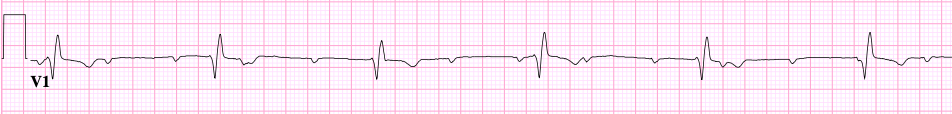
Second degree AV block — Mobitz I (Wenckebach)
ECG features
- Progressive PR prolongation across consecutive beats
- Eventual non-conducted P
- The cycle restarts with a shorter PR
- Largest PR increment occurs between the first and second conducted beats (the “footprint”)
- Grouped beating with shortening RR intervals before the dropped beat
Level of block
- Almost always AV nodal
- Worsens with vagal tone, improves with atropine
- Often physiologic in young, athletic, or sleeping patients
Clinical implications
- Usually benign in the absence of structural heart disease
- Can be a sign of inferior MI (RCA territory, AVN ischemia) — typically resolves
- Drug-related Wenckebach is common with beta-blockers, calcium channel blockers, digoxin
Pacing decision
- Not indicated if asymptomatic
- Class IIa for symptomatic Mobitz I or when block is infranodal (documented in the lab)
- Address reversible drivers first
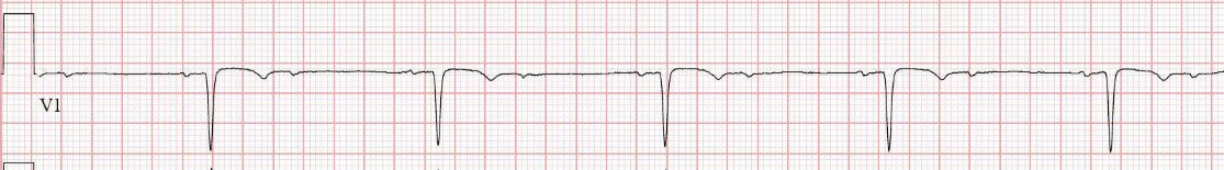
Second degree AV block — Mobitz II
ECG features
- Constant PR before the dropped beat
- Sudden failure of conduction with no warning
- Often wide QRS reflecting underlying BBB
- Grouped beating without progressive PR change
Level of block
- Infranodal (His or below)
- Does NOT improve with atropine; can worsen
- Vagal maneuvers do not modulate it
Clinical implications
- High risk of progression to complete heart block
- Syncope is the feared presentation
- Almost never benign — always investigate
Pacing decision
- Class I for symptomatic Mobitz II
- Class I for asymptomatic Mobitz II if infranodal — most cases qualify
- Pace early; don’t wait for syncope
High-grade / 2:1 AV block
ECG features
- ≥2 consecutive non-conducted Ps with preserved sinus rhythm
- 2:1 block is its own diagnostic puzzle — cannot distinguish Mobitz I from II on a single 12-lead because there’s no consecutive PR to compare
Localizing 2:1 block
- Narrow QRS + improvement with atropine → AVN
- Wide QRS + no atropine response → infranodal
- Carotid massage worsens AVN block, improves infranodal block (paradoxical)
- Exercise: AVN block improves with exercise; infranodal block worsens
- His bundle recording is definitive
Clinical implications
- Often symptomatic with bradycardia, fatigue, near-syncope
- Treat as Mobitz II until proven AV nodal
Pacing decision
- Class I for symptomatic high-grade block
- Class I for infranodal high-grade block regardless of symptoms
- Reversible causes must be excluded
Third degree (complete) AV block
ECG features
- Complete AV dissociation
- Atrial rate (regular Ps) faster than ventricular rate (regular escape)
- P waves “march through” the QRS at their own rate
- Escape rhythm regular at a slower rate than atrial
Escape morphology localizes
- Junctional escape, narrow QRS, 40–60 bpm: block within or just below AVN
- Ventricular escape, wide QRS, 20–40 bpm: infranodal block
- Lower escape = higher risk of asystole
Clinical implications
- Stokes-Adams syncope, dizziness, heart failure
- High risk of asystole, especially if escape is unreliable
- Investigate reversible causes urgently — MI, drugs, hyperkalemia, Lyme, endocarditis with aortic root abscess
Pacing decision
- Class I for symptomatic third-degree block
- Class I for asymptomatic third-degree if escape <40 bpm, exercise-induced, or with ventricular pauses
- Temporary pacing as a bridge to permanent device or to recovery from a reversible cause